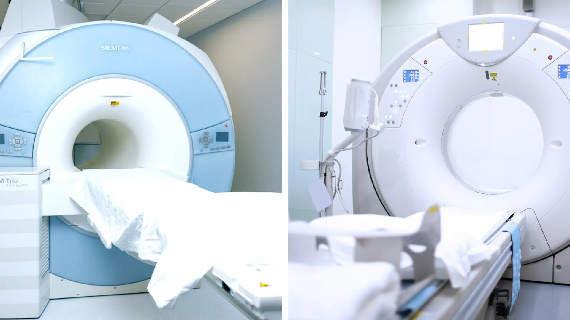
Magnetic Resonance Imaging
vs.
Computed Tomography
The differences between MRI and CT
Both Computed Magnetic Resonance Imaging (MRI) and Computed Tomography (CT) are sophisticated, high-resolution imaging technologies that have become increasingly relevant in modern medical practice. While they may seem similar, they have different uses and benefits. Let's explore these technologies from the ground up, outlining their uses, benefits, and potential risks to help clarify their roles in healthcare.
What is imaging technology?
Imaging technology refers to methods and techniques that allow doctors to see inside your body in a non-invasive way. This technology is used across a wide spectrum of medical fields for various purposes such as:
- Diagnosis: Imaging is often the first step doctors use in diagnosing many diseases and conditions by visualizing changes in the appearance of organs, tissues, and bones.
- Treatment planning: Imaging helps in planning surgical operations or other treatments, allowing precise treatments targeted to affected areas.
- Monitoring: Imaging is used to monitor the progress of diseases, the effectiveness of treatments, and to detect recurrence of illness.
- Research: Advanced imaging techniques are crucial in medical research, helping scientists and doctors better understand complex diseases .
Among the range of imaging tools available, MRI and CT stand out as particularly advanced, each using different technology suited for different medical uses. But what exactly are the differences between these two methods?
Understanding these distinctions is key to knowing when to use each type of scan. For instance, when is the fast imaging of a CT scan most necessary? Why might an MRI be a better option for detailed and repeated examinations? Answering these questions can help clarify how each technology fits with the specific need.
The technology behind MRI and CT scanners
Magnetic Resonance Imaging (MRI) uses a magnetic field along with radio waves rather than radiation to generate images. For an MRI, you lie on a table that gently slides you inside a large cylindrical device that is essentially a large magnet. The machine uses these magnetic and radio wave signals rather than radiation to produce detailed images of your organs and tissues, which are captured and processed into visual data. MRI is renowned for its exceptional ability to image soft tissues with high detail, and is particularly useful for in-depth studies of soft tissue-related injuries and diseases. It is also considered safer for repeated use and for sensitive groups of people.
Computed Tomography (CT) scans use X-rays to create detailed pictures of the inside of your body. During the scan, you lie on a table that slides into a large, doughnut-shaped machine. This machine takes X-rays from many different angles as it rotates around you. A computer then processes these X-rays to produce cross-sectional images, or slices, of your body, which can be combined to create a three-dimensional view. CT scans are useful for their fast imaging capabilities, often taking just a few minutes to complete. This speed is especially important in emergency situations where quick decisions need to be made, such as evaluating injuries from accidents or checking for severe internal problems.
What factors influence the choice between a MRI scan and an CT scan?
Choosing the right scan, whether it's a MRI or CT, mainly depends on what medical issue needs to be looked at. It also depends on what equipment is available. CT scans are often used simply because they are easier to find and can be used quicker, especially in emergencies or in places with fewer resources. However, this doesn't mean it's always the best option.
For instance:
- In urgent situations involving the brain, such as a suspected stroke or a severe head injury, a CT scan is often used because it quickly provides clear images to detect bleeding or fractures. However, when doctors need a closer look at brain tissue to diagnose conditions like tumors or chronic illnesses such as multiple sclerosis, an MRI is better.
- Similarly, with heart issues, doctors might use a CT scan to check for blockages in the coronary arteries or assess heart disease risk with calcium scoring. But, if they need detailed images of the heart's structure and function—such as evaluating the heart muscle's performance, examining complex heart conditions present from birth, or visualizing the blood vessels around the heart—an MRI is preferred, providing a fuller picture of cardiovascular health.
- When checking for complex bone fractures, CT scans are usually the best choice because they show detailed images of the bones. This is really helpful for seeing small pieces of the bone and figuring out exactly how the bone is broken, since bones are hard and show up well on CT scans. On the other hand, MRI is better for looking in general at the bones, bone marrow, and the soft tissues around the bones because it gives a clearer view of these softer areas.
- Certain organs, such as the lungs, are more effectively visualized using a CT scan, whereas an MRI excels in imaging various other organs like the brain, liver, and kidneys.
For more in-depth information on the many uses of MRI click here.
How does the image quality and detail compare?
Both CT and MRI scans offer high-resolution images, but the type of detail and what is emphasized can differ greatly between the two:
CT Scan: Offer excellent clarity for solid structures, such as bones, making them ideal for evaluating skeletal issues. They can also visualize changes in soft tissues and blood vessels fairly well. However, when it comes to the fine details in soft tissue, CT scans are generally less effective than MRI. To match the detail of an MRI, a CT scan would need to use a contrast agent.
MRI Scan: Is superior in terms of soft tissue distinction. It can clearly differentiate between the various types of soft tissues, which is crucial for accurately diagnosing issues within the brain, muscles, heart, and identifying soft tissue cancers.
What are the specific risks associated with each type of imaging technology?
CT Scan: The primary risk involved with CT scans is their use of ionizing radiation. Regular exposure to this radiation can be concerning for children or individuals who require frequent scans, as it could potentially impact long-term cellular health. Additionally, CT scans more often require the use of contrast to improve the clarity of the images of internal structures. While this is generally safe, they can trigger allergic reactions in some patients.
MRI Scan: A significant advantage of MRI is that it does not use ionizing radiation, making it the safer option for patients especially those needing multiple scans, like those managing chronic conditions or under long-term medical observation. The main risk with MRI involves patients with specific metal implants, like pacemakers or some surgical clips. These implants may interfere with the MRI's magnetic fields, and could potentially affect the device and quality of the scan. Many implants nowadays are MRI compatible, but it's always important to check this before booking a scan. While MRIs also sometimes use contrast agents to enhance image quality, it is required less frequently than with CT scans.
Visit our FAQ page for more information on MRI safety.
Conclusion
Choosing between a CT scan and an MRI is a decision that depends heavily on the specific medical circumstances, the particular area of the body being examined, and the patient's comprehensive medical history. Both technologies offer distinct advantages and have their own limitations, making them invaluable in their respective capacities for diagnosing and monitoring various health conditions.
Carefully choosing the right imaging method ensures patients get accurate diagnoses and personalized treatment plans for their health issues. Using these advanced imaging technologies thoughtfully improves patient outcomes, helps manage treatment effectively, and enhances the overall quality of healthcare services.

